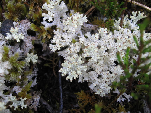
About Pulchrocladia retipora (Labill.) S.Stenroos, Pino-Bodas & Ahti
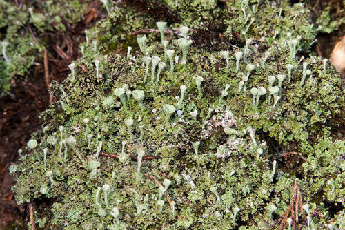
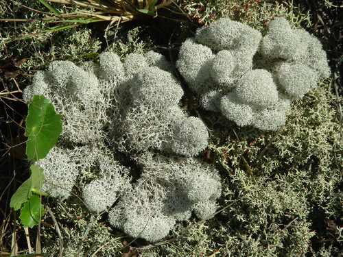
Like most species in the Cladoniaceae family, Pulchrocladia retipora has a cladoniiform growth form, meaning it produces both a primary horizontal thallus and a secondary vertical thallus. The primary thallus is white, nodular (marked by small raised areas or swellings), and is short-lived. Podetia, the secondary thallus, grow from the primary thallus. They typically reach up to 5 cm (2 in) tall, and are coloured white to pale grey, sometimes with a pinkish tinge, yellowing, or superficial blackening at their tips. Podetia are rigid when dry, and become spongy when wet. They branch irregularly or dichotomously, forming cushiony clumps. Their wall is highly perforated, with around 5–11 large, round to ellipsoidal holes per centimetre. The podetium surface is continuously corticate and lacks soredia. The inner medulla is formed of twisted hyphal strands with a cobweb-like structure. Even though it lacks surface soredia, P. retipora produces soredia-like clusters of algal cells and hyphae within the inner medulla strands.
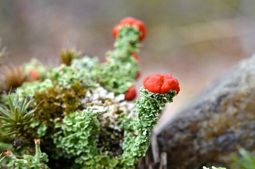
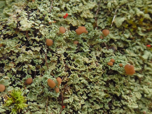
Apothecia (sexual reproductive structures) develop infrequently. When present, they are small, black, peltate, and grow crowded together at the ends of small terminal branchlets. The hymenium ranges in colour from dark reddish-brown to black. Ascospores measure 25–27 by 5 μm. Conidiomata form at the ends of branchlets, are covered in translucent slime, and produce curved or straight conidia that measure 6 by 1 μm. Collected specimens from Australia tend to have brownish colouration, while specimens from New Zealand vary from pure white to grey, grayish-green, or slightly yellowish. The photobiont (photosynthetic partner) of this lichen is a green alga from the genus Trebouxia; occasionally free-living algae become trapped in the irregularly intertwined hyphae of the medulla.
The major secondary compounds found in P. retipora are usnic acid and atranorin. Smaller quantities of other compounds, identifiable via thin-layer chromatography, include protolichesterinic acid, ursolic acid, and in most cases rangiformic acid and norrangiformic acid. Results from in vitro experiments suggest usnic acid is responsible for the antimicrobial, antiviral, and cytotoxic biological activity seen in P. retipora extracts. The concentration of usnic acid in the thallus determines the colour of P. retipora, creating a colour range from opaque greyish-white through yellowish-white to a distinct yellow.
Pulchrocladia retipora is closely related to P. corallaizon, and the two species are often confused due to their similar appearance. They can be distinguished by the inner medulla structure: P. retipora has a characteristically tightly packed inner medulla. More mature pseudopodetia of C. corallaizon may have regions where the inner medulla is missing or less dense, though the medulla stays compact in the top branches of pseudopodetia, so it never appears stranded or corticated.
Pulchrocladia retipora, also called coral lichen, is widely distributed across Australasia. In Australia, it has been recorded in the Australian Capital Territory, New South Wales, Queensland, Victoria, and Tasmania. In New Zealand, it occurs on the North Island, South Island, Antipodes Islands, Auckland Islands, Campbell Island, and Chatham Islands. Outside of Australasia, it grows in New Caledonia in the Pacific.
This lichen is common in subalpine peat bogs, and often grows in association with the lichens Cladonia confusa, Rexiella sullivani, and Stereocaulon ramulosum. It grows on peaty soil among tussocks, or in heaths dominated by Dracophyllum and Leptospermum, most frequently at the margins of Nothofagus forests and in fellfield; it rarely grows on other substrates such as rocks, logs, and sand dunes. In the moorlands of Tasmania's Meredith Range, it grows well in well-drained, elevated locations, especially near decomposing buttongrass hummocks.
P. retipora reproduces vegetatively: new podetia grow from fragments of old podetia. Its growth rate is highly variable, ranging from less than 1 mm per year to up to a few centimetres per year. It grows in clusters that can sometimes reach one metre in diameter. Its cushion-like growths range in diameter from roughly 10 cm (4 in) to 100 cm (40 in). New Zealand botanist William Martin recorded finding square metre-sized cushions in the Lewis Pass area of Canterbury, New Zealand. Football-sized lichen cushions have been observed on the mountain range in Australia's Grampians National Park. Martin noted that large growths only form in subalpine zones; lowland growths reach just 5–10 centimetres (2–4 in) in size. The lichen's unique morphology helps it survive in exposed heath habitats: its net-like coral structure increases gas exchange, moderates temperature extremes, and maximizes access to light and water.
The complex net-like structures of P. retipora are called fenestrations. Lichenologist Rosmarie Honegger described the thallus of this species as "likely to be among the most complex vegetative structures ever produced in the fungal kingdom". The lichen's appearance has been described as "of considerable beauty resembling lace or coral". Because of this attractive appearance, it has been used in floral decoration and architectural design. German lichenologist Robert Lücking highlighted the cover design of volume 37, issue 1 of *The Lichenologist* (published in 2005) as particularly striking in his review of the journal's cover designs; this issue featured an image of P. retipora set against a blue-themed background.