About Coprinellus micaceus (Bull.) Vilgalys, Hopple & Jacq.Johnson
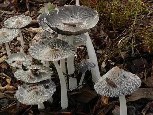
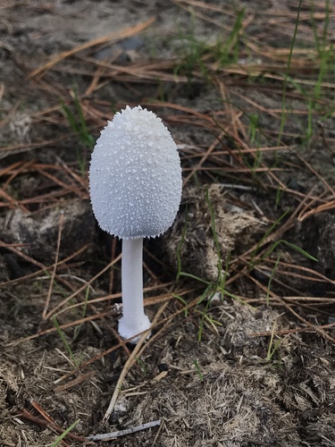
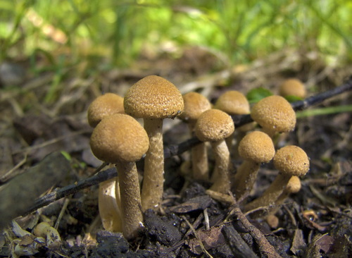
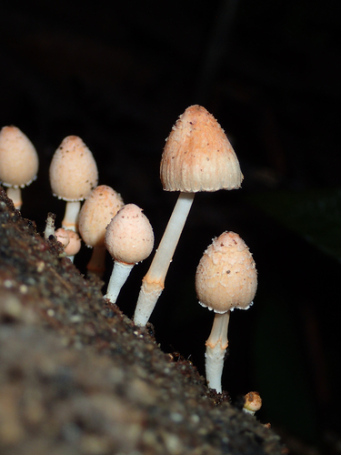
Coprinellus micaceus (Bull.) Vilgalys, Hopple & Jacq.Johnson has the following macroscopic characteristics. When immature, its cap is 1–2.5 cm (1⁄2–1 in) in diameter, with an oval to cylindrical shape. As it matures, the cap expands to a bell (campanulate) shape, sometimes with a central nipple-like protrusion called an umbo, before finally becoming slightly flattened and convex. When fully expanded, the cap reaches 0.8–5 cm (1⁄4–2 in) in diameter; its margin tears into rays and curves slightly upward. The cap is yellow-brown or tan, often with a darker center, fading to pale yellow or buff moving inward from the margin. The cap margin has prominent grooves that extend almost to the cap center, marking the position of longer gills on the cap's underside. Young specimens have a cap surface covered in loose, white or whitish shiny particles, which are remnants of the universal veil that covers immature fruit bodies. These particles wash away easily, so older specimens usually have smooth caps. This species is hygrophanous, meaning it changes color depending on its hydration level. Its gills are closely crowded, with a narrow (adnexed) attachment to the stipe. Gills start out white, then turn dark brown, and eventually become black as spores mature. Cap expansion splits gills open along their midline, which tears the cap margin into rays. Spore discharge and autodigestion start at the base of the gills, before the upper sections of the gills fully turn black. The stipe is brittle, hollow, and measures 3–10 cm (1+1⁄4–4 in) long by 2–5 mm (1⁄16–3⁄16 in) thick, with roughly consistent diameter along its entire length. It is generally white, but may discolor to pale dirty cream starting from its base. Young stipes have a velvety surface covered in very fine whitish powder, which wears off over time to leave the stipe more or less smooth. A rudimentary ring, another remnant of the universal veil, may be present at the stipe base. The spore print of C. micaceus is dark brown or black. Its flesh is thin and fragile, white in the stipe and brownish in the cap. It has no distinctive odor or taste. Individual fruit bodies take an average of five to seven days to reach full maturity. For microscopic characteristics, C. micaceus spores are reddish-brown or black, with dimensions of 7–10 by 4.5–6 μm. They are typically biconvex lens-shaped (lentiform): when viewed from the side, they look more almond-shaped or spindle-shaped, while in front view they look oval or mitriform (roughly the shape of a peaked miter cap). Spores have a germ pore, a flattened central area on the spore surface through which a germ tube can emerge. The spore-bearing basidia are four-spored, club-shaped, and measure 10–15 by 4–7 μm. Studies show basidia develop in four distinct generations. First-generation basidia are the most protrusive and extend the farthest from the hymenium surface. Subsequent generations of basidia are shorter and less protrusive. Four distinct sets of basidia can be seen clearly when a living gill is viewed under a microscope. Arthur Buller created the term inaequihymeniiferous to describe this hymenial development pattern. Staggered basidia sizes are arranged this way to make spore release from the hymenium easier. There are four spore discharge zones that correspond to the four sets of basidia, and basidia that have released all their spores quickly begin to autodigest. This staggered arrangement minimizes the chance of spores colliding with neighboring basidia during release. Cheilocystidia (cystidia located along the cap edge) are spherical, and measure 30–120 by 20–74 μm. Pleurocystidia (facial cystidia on the gill face) are club-shaped or elongated ellipses, reaching up to 130–155 μm in length. Pleurocystidia protrude from the gill face and act as spacers, preventing adjacent gills from touching and ensuring enough space for basidia and spore development. C. micaceus may also have scattered caulocystidia (cystidia on the stipe) measuring 60–100 by 5–10 μm, but their presence is variable and cannot be used for reliable identification. Both De Bary and Buller, in their studies of cystidia structure, concluded that a central mass of cytoplasm forms where numerous thin cytoplasmic plates meet at the cell's center. De Bary thought the plates were filamentous branching processes, while Buller proposed they form in a process similar to foam bubble walls, and that the central mass can slowly change shape and position by altering the relative volume of vacuoles enclosed by the thin cytoplasmic walls. In older cystidia cells, cytoplasm may be restricted to the cell periphery, with a single large vacuole occupying the cell center. The globular cells that form the mica-like scales on the cap are colorless with smooth walls, and range in size from roughly 25–65 μm, with most falling between 40 and 50 μm. Buller described the glitter of these cells: "The sparkling of the meal-cells, as well as of the cystidia on the edges and faces of the gills, is simply due to light which strikes them from without and is refracted and reflected to the eye in the same manner as from the minute drops of water one so often sees at the tips of grass leaves on English lawns early in the morning after a dewy night." In 1914, Michael Levine became the first person to report successfully cultivating C. micaceus from spores in a laboratory setting. In his experiments, fruit bodies appeared around 40 to 60 days after spores were inoculated into growth media (agar supplemented with soil, horse dung, or cornmeal). Like other coprinoid species, C. micaceus undergoes synchronous meiosis. Its chromosomes are easily visible with light microscopy, and all meiotic stages are well-defined. These features have made the species a useful tool for laboratory studies of basidiomycete cytogenetics. The haploid chromosome number of C. micaceus is n=12. Because its microscopic characteristics and cytogenetics are well-documented, the species has frequently been used as a model organism to study cell division and meiosis in basidiomycetes. In terms of ecology, habitat, and distribution, C. micaceus is a saprotrophic species that gets nutrients from dead, decomposing organic matter. It grows in and around stumps or logs of broad-leaved trees, or attached to buried wood. It prefers feeding on bark, particularly secondary phloem, over wood itself. In the successional sequence of fungal species that decompose wood, C. micaceus is a late-stage colonizer that prefers wood that has already decomposed enough to become friable and softened. A 2010 study suggests the fungus can also live as an endophyte, growing in the woody tissue of healthy trees without causing disease symptoms. It is also associated with disturbed or developed ground, including roadsides, path edges, gardens, construction sites, and parking lot edges. It has also been recorded growing indoors on rotting wood in humid environments, and was once found growing on wooden roof support gangways and props around 120 m (400 ft) underground in an abandoned coal mine. Fruit bodies commonly grow in dense clusters, but can also grow singly or in small clumps, especially in forested areas. In North America, C. micaceus is one of the first edible mushrooms to appear in spring, and fruits from April to September. In Europe, it fruits from May to December. While it can grow at any time of year, it is most common in spring and fall, which coincides with higher humidity from spring and autumn rains. An air quality study conducted in Santiago de Compostela, Iberian Peninsula, found that most "Coprinus" spores in the atmosphere belonged to C. micaceus, and that spore counts increased with higher humidity and rainfall, but decreased at higher temperatures. The species is known for producing successive crops of fruit bodies at the same location. In one recorded case, a total of 38 lb (17.2 kg) of fresh mushrooms were collected from a single elm stump across 10 successive crops over one spring and summer. C. micaceus has a cosmopolitan distribution, and has been collected in northern Africa, South Africa, Europe (including Turkey), North America (as far north as Alaska), the Hawaiian islands, South America, India, Australia, New Zealand, and Japan. Phylogenetic analysis of rDNA sequences from specimens collected in southeastern Asia and Hawaii shows that Hawaiian populations form a distinct clade with low genetic diversity compared to Asian populations, which suggests Hawaiian populations were introduced relatively recently and have not had enough time to accumulate significant genetic variation. One study suggests that in South Africa, where C. micaceus is rare, it has often been mistaken for the similar-looking, more common species C. truncorum. A similar observation has been made about North American populations.