About Amanita gemmata (Fr.) Bertill.
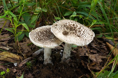
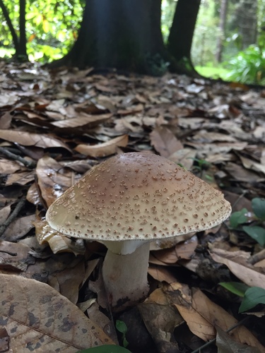
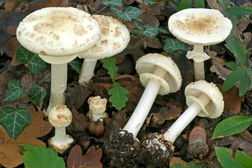
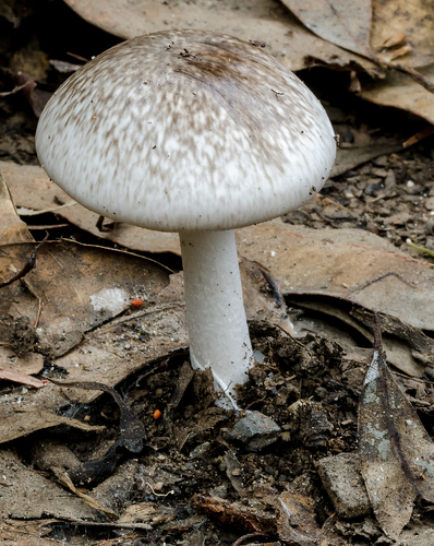
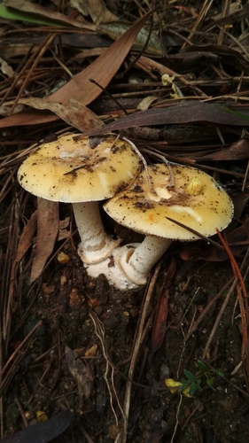
Amanita gemmata (Fr.) Bertill. has fruit bodies that are yellow overall. Fresh caps range in color from dull creamy yellow to golden yellow to buff, and are sticky when moist. The cap surface is covered in randomly distributed white warts that are usually flimsy and easily washed away by rain, though the warts tend to be more concentrated toward the cap center. Mature caps are typically 2.5–12 centimetres (1–4+3⁄4 inches) in diameter, starting out convex before flattening out as they mature. The flesh is white and does not change color when sliced. Gills are adnate to adnexed, white, and closely spaced with very little space between them. The pale yellowish stem is 4–12 cm (1+5⁄8–4+3⁄4 in) long by 0.5–1.9 cm (1⁄4–3⁄4 in) thick, and is either roughly consistent in width along its length, or slightly thicker at the base. Young mushrooms have a membranous partial veil that stretches from the upper stem to the cap margin; as the mushroom matures, this veil tears and leaves a flimsy, skirt-like, easily lost ring on the stem. At the base of the stem is a white volva, a remnant of the universal veil that covered the immature mushroom, which usually forms a small, free rim. The spore print is white, and there is no distinctive odour. Microscopically, Amanita gemmata has ellipsoid to broadly ellipsoid spores that measure 8–10 by 6.5–7.5 μm, with an average length-to-width Q-ratio of 1.35. Spores are not amyloid, and are smooth, thin-walled, and contain one to several small oil droplets. The spore-bearing basidia of the hymenium are usually four-spored, club-shaped, and measure 30–40 by 8–11 μm. Gill tissue is divergent, meaning cells are roughly parallel near the center of the gill, but bend outward toward the gill edge. Hyphae in gill tissue are cylindrical to inflated, thin-walled, translucent (hyaline) to yellowish, and measure 2.2–9 μm wide; hyphae in the central strand of the gill are narrower and typically cylindrical. Hyphae of the subhymenium, the tissue layer directly beneath the hymenium, are interwoven, branched, cylindrical to slightly inflated, hyaline, and 6–9 μm wide. Hyphae of the cap cuticle are filamentous, interwoven, radially arranged, cylindrical, 2.7–4 μm wide, thin-walled, hyaline to yellowish, and gelatinize when mounted in potassium hydroxide. Cap tissue is also interwoven, with hyphae that are cylindrical to somewhat inflated, 3.7–14.6 μm wide, thin-walled, branched, and hyaline to yellowish. Caulocystidia are abundant on the apex of the stem; they are club-shaped to cylindrical, thin-walled, hyaline, and measure 3–9 μm wide. Annulus tissue is made up of interwoven cylindrical hyphae measuring 3–9 μm wide, and also contains inflated spherical sphaerocysts that are club-shaped to ellipsoidal, with dimensions of 29–55 by 30–70 μm. The cap warts, which are remnants of the universal veil, are made of loosely interwoven, cylindrical to inflated, thin-walled hyphae that are 3.5–8 μm wide. Sphaerocysts in the cap wart tissue are 58.5–70.2 by 17.5–40 μm, ellipsoidal, and hyaline. Volval tissue is interwoven, with cylindrical, hyaline hyphae that are 4.4–7.3 μm wide. Sphaerocysts in volval tissue are ellipsoidal to roughly spherical, hyaline, and measure 35–70 by 20–35 μm. In A. gemmata, refractive cells are scattered, most abundant just below the cap cuticle, and measure 3.7–6 μm wide. Clamp connections are rare in the hyphae of A. gemmata, but are present in the annulus, gill tissue, subhymenium, and cap tissue. This fungus prefers sandy and slightly acidic soils, and is often found growing in association with Norway Spruce (Picea abies). It grows singly, scattered, or in groups in coniferous and mixed forests, especially along paths and roads. The species is distributed across parts of Asia and Europe. As a species cluster, it is widely distributed in North America, where it has been found as far south as Ixtlán de Juárez, Mexico. It has also been reported from the Dominican Republic, and in South America it is known from Chile and Colombia. In Asia, it has been collected from Iran and China. Amanita gemmata is a mycorrhizal fungus, meaning it forms a mutually beneficial relationship with the roots of compatible host plants. Through this association, the plant provides the fungus with a carbon source, and the fungus provides the plant with benefits including nutrients and protection from pathogens. Largent and collaborators (1980) documented mycorrhizal associations of A. gemmata with Manzanita (Arctostaphylos spp.) and Pinus contorta (lodgepole pine), and Nieto and Carbone documented an association with Pinus pinaster (maritime pine) in Spain. Toxicity is suspected to be caused by muscimol and ibotenic acid, compounds also found in many species in Amanita section Amanita, including A. muscaria and A. pantherina. Poisoning symptoms generally appear within three hours of ingestion, and include visual hallucinations, nausea, vomiting, stomach pain, diarrhea, irregular slow heartbeat, and agitation. Severe cases involving coma, convulsions, or death are extremely rare.