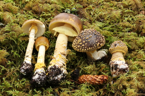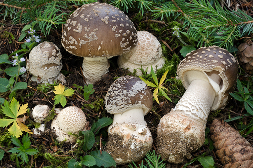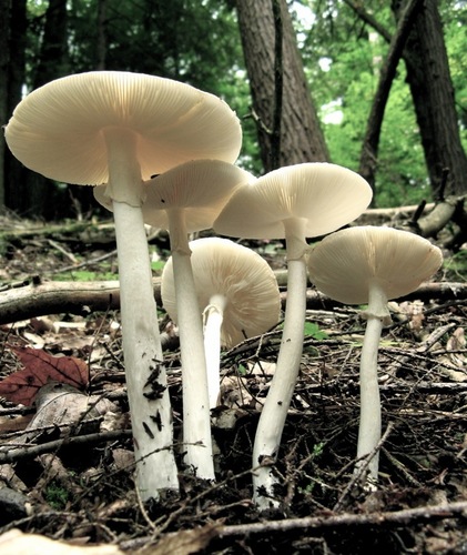
About Amanita bisporigera G.F.Atk.
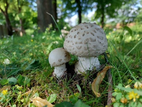
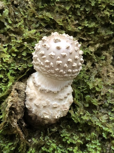
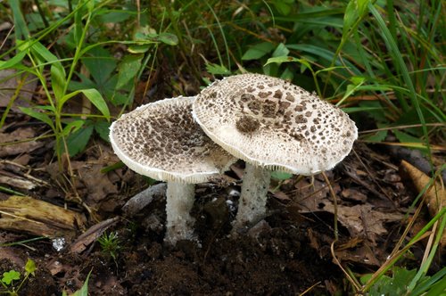
The cap of Amanita bisporigera is 3–13 centimeters (1–5 inches) wide. Its shape changes with age, shifting from egg-shaped to convex to somewhat flattened. The cap surface is smooth and white, sometimes with a pale tan or cream tint in the center. It is either dry, or slightly sticky when environmental conditions are moist. The mushroom's flesh is thin, white, and does not change color when bruised. In young specimens, the cap margin is rolled inwards, and it lacks striations (grooves) and volval remnants. The gills are also white, closely crowded, and either not attached to the stipe or only barely reach it. Lamellulae (short gills that do not extend all the way to the stipe) are numerous and narrow gradually. The white stipe measures 6–14 cm (2+1⁄2–5+1⁄2 in) long by 0.7–1.8 cm (1⁄4–3⁄4 in) thick. It is solid (not hollow), tapers slightly upward, and its surface is frequently floccose (covered with tufts of soft hair), fibrillose (covered with small slender fibers), or squamulose (covered with small scales), especially in young specimens; fine grooves may run along its length. The bulb at the stipe base is spherical or nearly spherical. A delicate white, thin, membranous skirt-like ring sits on the upper stipe; this is a remnant of the partial veil, which extends from the cap margin to the stalk and covers developing gills. When young, the entire mushroom is enveloped in a universal veil membrane that stretches from the top of the cap to the bottom of the stipe, giving it an oval, egg-like shape. In mature fruit bodies, remnants of this veil form an eggshell-shaped cup-like volva around the mushroom base. Occasionally, the volva stays underground or tears apart during development. It is white, sometimes lobed, and may lie pressed closely against the stipe. Measured from the base of the bulb, the volva reaches up to 3.8 cm (1+1⁄2 in) in height, and is about 2 mm thick midway between its top and base attachment. The mushroom's odor has been described as "pleasant to somewhat nauseous", becoming more cloying as the fruit body ages. When a 5–10% potassium hydroxide (KOH) solution (a common mushroom identification chemical test) is applied to the cap flesh, it turns yellow. A. bisporigera shares this characteristic chemical reaction with A. ocreata and A. virosa, though some authors have questioned the identity of North American A. virosa, suggesting these collections may actually be four-spored A. bisporigera. Tulloss suggests that reports of A. bisporigera that do not turn yellow with KOH were actually based on white forms of A. phalloides. Collections from the Chiricahua Mountains of Arizona and central Mexico, while nearly identical in appearance to A. bisporigera, do not stain yellow with KOH; their taxonomic status has not been investigated in detail. Like most other Amanita species, A. bisporigera is thought to form mycorrhizal relationships with trees. This mutually beneficial relationship sees the fungus' hyphae grow around tree roots, allowing the fungus to receive moisture, protection, and nutritive byproducts from the tree, while giving the tree greater access to soil nutrients. A. bisporigera fruit bodies grow on the ground, either solitarily, scattered, or in groups in mixed coniferous and deciduous forests, and tend to appear during summer and early fall. They are commonly found near oak, but have also been reported in birch-aspen areas in western North America. The species is most common in eastern North America, and rare in western North America. It is widely distributed in Canada, and its range extends south to Mexico. It has also been found in Colombia, where it may have been introduced via trees exported for use in pine plantations. A. bisporigera is considered the most toxic North Amanita mushroom, with little variation in toxin content between different fruit bodies. Three subtypes of amatoxin have been identified: α-, β-, and γ-amanitin. The main amatoxin, α-amanitin, is readily absorbed through the intestine; 60% of the absorbed toxin is excreted into bile and undergoes enterohepatic circulation, while the kidneys clear the remaining 40%. The toxin inhibits the enzyme RNA polymerase II, which interferes with DNA transcription, and suppresses RNA production and protein synthesis. This causes cellular necrosis, especially in cells that are initially exposed and have rapid rates of protein synthesis, leading to severe acute liver dysfunction and ultimately liver failure. Amatoxins are not broken down by boiling, freezing, or drying. Roughly 0.2 to 0.4 milligrams of α-amanitin is present in 1 gram of A. bisporigera, and the lethal dose for humans is less than 0.1 mg per kg of body weight. One mature fruit body can contain 10–12 mg of α-amanitin, enough for a lethal dose. The α-amanitin concentration in the spores is about 17% of the concentration found in fruit body tissues. A. bisporigera also contains phallacidin, a phallotoxin structurally related to amatoxins but considered less poisonous due to poor absorption. Poisonings from similar white amanitas have been reported in domestic animals including dogs, cats, and cows. The first reported fatal poisonings from consumption of A. bisporigera occurred near San Antonio, Mexico, in 1957: a rancher, his wife, and three children ate the fungus, and only the man survived. Amanita poisoning has three distinct stages. First is the incubation stage, an asymptomatic period that lasts 6 to 12 hours after ingestion. Next is the gastrointestinal stage, which starts around 6 to 16 hours after ingestion: abdominal pain, explosive vomiting, and diarrhea develop and last up to 24 hours, which can lead to dehydration, severe electrolyte imbalances, and shock. These early symptoms are possibly linked to other toxins such as phalloidin. The cytotoxic stage occurs 24 to 48 hours after ingestion: clinical and biochemical signs of liver damage appear, while patients typically no longer have gastrointestinal symptoms. Signs of liver dysfunction include jaundice, hypoglycemia, acidosis, and hemorrhage. Later, prothrombin and blood ammonia levels increase, and signs of hepatic encephalopathy and/or kidney failure develop. Reported risk factors for death include age younger than 10 years, a short latency period between ingestion and symptom onset, severe coagulopathy (blood clotting disorder), severe hyperbilirubinemia (jaundice), and rising serum creatinine levels.